|
Product Details:
|
| Product Name: | Pepsinogen I / Pepsinogen II (PGI / PGII) Combo Test Kit (TRFIA) | Funtion: | Gastric Function Diagnose |
|---|---|---|---|
| Technology: | Time Resolved Fluorescence Immunochromatographic Assay (TRFIA) | Usage: | Vitro Diagnostic Reagent |
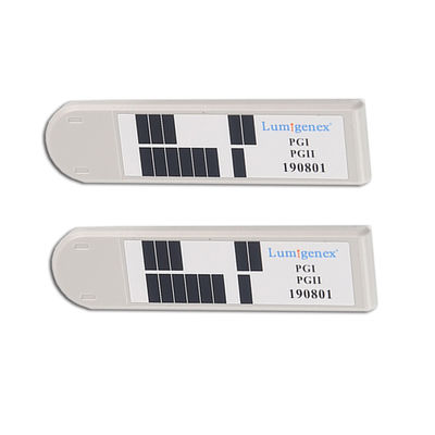
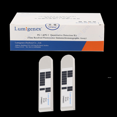
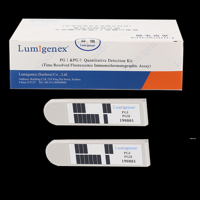
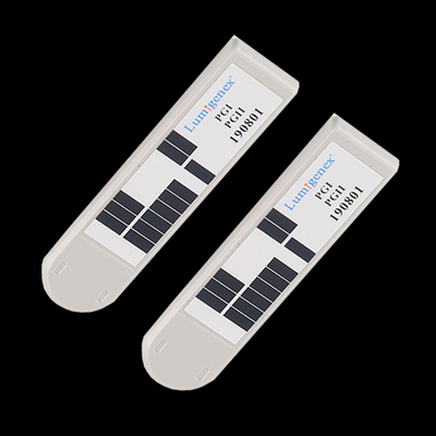
| Format: | Strip, Cassette | Specimen: | Whole Blood, Plasma, Serum |
| Storing Temperature: | 2℃-8℃ | Shelf Life: | 18 Months |
| Reading Time: | 15 Min. | Compatible Equipment: | Lumigenex TRFIA Analyzer LTRIC-600, LTRIC-1000 |
| Highlight: | poc rapid strep test,poc kits |
||
High sensitivity cFDA approved Pepsinogen I / Pepsinogen II (PGI / PGII) Combo Test Kit (TRFIA)
The combined detection kit of pepsinogen I/ pepsinogen II (time-resolved fluorescence immunoassay) is suitable for quantitatively determining pepsinogen I, pepsinogen in human serum, plasma, and whole blood in vitro.
The concentration and PGI/PGII ratio of pepsinogen I (hereinafter abbreviated as PGI), pepsinogen II (hereinafter abbreviated as PGII) can be used in the diagnosis of atrophic diseases of gastric fundus mucosa, such as superficial gastritis, erosive gastritis, gastric ulcer, duodenal ulcer, atrophic gastritis, etc. Pepsinogen (PG) is an inactive precursor of pepsin (proteolytic enzyme) in gastric juice, Immunology can be divided into two types: pepsinogen I (PG I) and pepsinogen II (PG II). PG I is secreted by the stomach glands, PG II is secreted by the gastric fundus gland, cardia gland, pyloric gland, and Brunner gland. In the process of gastric mucosal atrophy, A decrease in the number of cells that secrete PG I, Hyloric gland cell proliferation, As a result, the PGI/PGII ratio is reduced.
Principle
The pepsinogenI/ pepsinogen II combined detection kit (time-resolved fluorescence immunochromatography) was used to quantitatively determine the concentration of pepsinogen I, pepsinogen II in serum, plasma and whole blood based on Time Resolved Fluorescence Immunochromatographic Assay.
The sample detection solution and blood samples were mixed and added to the reagent card. The sample (PGI/PGII) was combined with the mouse anti-human pepsinogen I monoclonal antibody microsphere fluorescent probe, mouse anti-human pepsinogen II monoclonal antibody nano-microsphere fluorescent probe on the marker pad to form a complex and the monoclonal antibody containing mouse anti-human pepsinogen I, mouse anti-human pepsinogen II was detected on the nitrocellulose membrane by capillary action chromatography and captured, Form a double antibody sandwich complex (that is, monoclonal antibody-to-test-mab nano-microsphere fluorescent probe complex). As a result, the more PG, PGII in the sample, the more the double antibody sandwich complex accumulates. The excess unreacted Mono-anti-nano-microspheres fluorescence probe continued to chromatography to the control line. The fluorescence intensity on the detection line is positively correlated with the concentration of the object (PGI, PGII) in the sample, and the regression equation is calculated and written into the IC card.
The Time-Resolved Fluorescence Immunoanalyzer reads the regression equation data in the IC card, tests the fluorescence intensity on the detection line, and then replaces the fluorescence intensity with the regression equation for automatic calculation, that is, the concentration of the object (PGI/PGII) in the output sample.

Recommended Products
![]()
|
Detection Projects
|
Test Strips and Cassettes (with links)
|
|
Diabetes |
HbA1C, Glucose, Ketone Body |
|
Hyperlipidemia |
Lipid Panel |
|
Anemia |
Hemoglobin |
|
Cardiac |
CK-MB, cTnI, Myo, NT-proBNP, D-Dimer, H-FABP, sST2, Homocysteine, cTnI/H-FABP, cTnI/CK-MB/Myo |
|
Inflammation |
CRP, PCT, SAA, CRP/SAA, PCT/IL-6 |
|
Hormone |
5(OH)D3, β-HCG, AMH |
|
Gastric Function |
PGI/PGII |
|
Liver Injury |
Aspartate Transaminase, Alanine Aminotransferase |
|
Renal Injury |
mALB, Creatinine, NGAL, ACR(TRFIA), ACR(Colloidal Gold) |
|
Gout |
Uric Acid |
|
Others |
Influenza A+B Combo, Influenza A+B/RSV Combo Monkeypox CG, Monkeypox IgG/IgM, Monkeypox RT-PCR |
Contact Person: Bonnie
Tel: 86-13814877381


 English